Fig. 2.
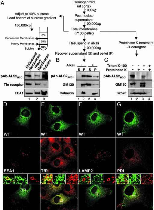
Endogenous WT ALS2 is a peripherally associated endosomal membrane protein oriented toward the cytoplasm. (A–C) Total membranes were processed for flotation centrifugation, alkali extraction, and proteinase sensitivity assays as illustrated. (A) Endogenous ALS2 is detected primarily in endosomal membrane fractions. After centrifugation, the membrane-containing interfaces and an aliquot of the layer containing soluble proteins (40%) were collected and immunoblotted as indicated. (B) Endogenous ALS2 is peripherally associated with intracellular membranes. After alkali extraction, aliquots of supernatants (S) and membrane-containing pellets (P) were immunoblotted as indicated. (C) Endogenous ALS2 resides on the cytoplasmic face of intracellular membranes. Aliquots of total membranes were treated with proteinase K in the presence or absence of detergent and immunoblotted as indicated. (D–G) Transfected ALS2 colocalizes with the endosomal markers EEA1 and transferrin receptor (TfR). Representative deconvolved images of COS cells transiently transfected with WT ALS2 (FITC, green, Top) and stained for endogenous membrane markers (Cy5, red, Middle): EEA1 (D), TfR (E), lysosome-associated membrane protein-2 (LAMP-2) (F), and protein disulfide isomerase (PDI) (G). Merged images together with enlarged boxed area that demonstrates merged (Left), ALS2 (green, Center), and endogenous marker (red, Right) images are shown (Bottom). (Magnifications: ×600.)

