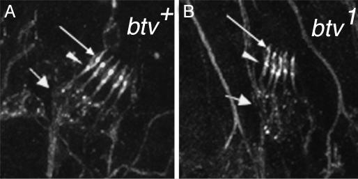
Fig. 2.
Morphological defects in cho mutants. Late-stage embryos were stained with α-HRP as detailed in Table 2. Shown are abdominal hemisegments of a control embryo (40AG13) (A) and a btv1 mutant embryo (B). The btv mutants showed a high frequency of outer dendritic segment defects, especially the lack of a clear ciliary dilation (thin arrow). The inner dendritic segments, from the soma (thick arrow) to the basal bodies (arrowhead), appeared relatively normal in btv mutants compared to the 40AG13 controls. We found no obvious defects in the 5D10 mutants (not shown; see also Table 2), which are the weakest of our cho mutants.

