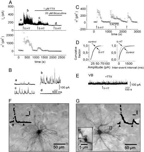
Fig. 1.
Effect of 5-HT receptor activation on IPSC activity in rat thalamocortical relay neurons. (A) Membrane current (Im) and membrane current variance (σ2) of a LGNd relay neuron during activation of 5-HT receptors (application of 5-HT at 10 μM for 1.5 min at times indicated by arrows) in normal ACSF and in the presence of 1 μM TTX and 20 μM bicuculline (indicated by bars). (B) Three-second intervals of the continuous current trace in A depicted at an expanded time scale at the times indicated (a-d). (C) Occlusion of the 5-HT-induced increase in IPSC activity during nearly maximal action of (±)-1-aminocyclopentane-trans-1,3-dicarboxylic acid (t-ACPD) (125 μM). TTX (1 μM) was present throughout the experiment. (D) Cumulative probability plots obtained under control conditions and in the presence of 10 μM 5-HT (synaptic events were counted over time periods of 3 min). Recordings were obtained in TTX (1 μM). (E) Membrane current of a ventrobasal complex (VB) relay neuron; 100 μM 5-HT was applied (for 1.5 min) at the time indicated by the arrow in the continuous presence of TTX (1 μM). (F and G) Photomicrographs of biocytin-labeled type I (F) and type II (G) neurons. Traces show responses of the same cells to 5-HT (application indicated by arrowheads) before (first response) and after (second response) addition of TTX. [Scale bars: 200 s, 200 pA2 (F); and 200 s, 400 pA2 (G).] The neuron in F displays radial dendritic morphology consistent with type I morphology and lacks TTX-insensitive responses to 5-HT. The neuron in G possesses numerous swellings along dendrites and near branch points typical of type II morphology (Inset) and generates TTX-insensitive responses to 5-HT. Note the existence of TTX-sensitive responses in both types of neuron.

