
Figure 2.
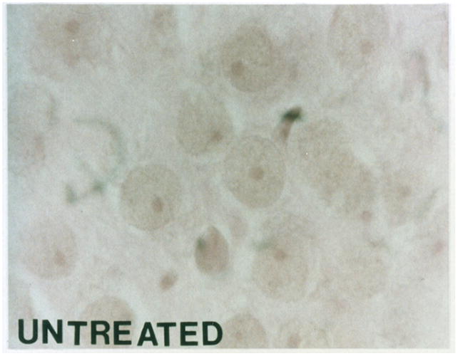
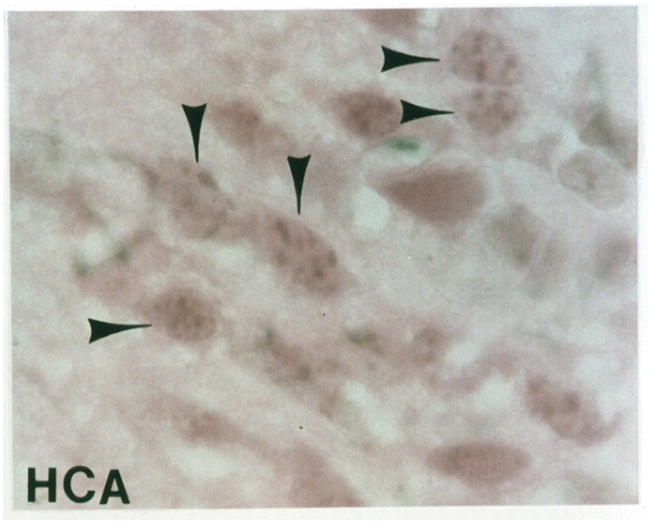
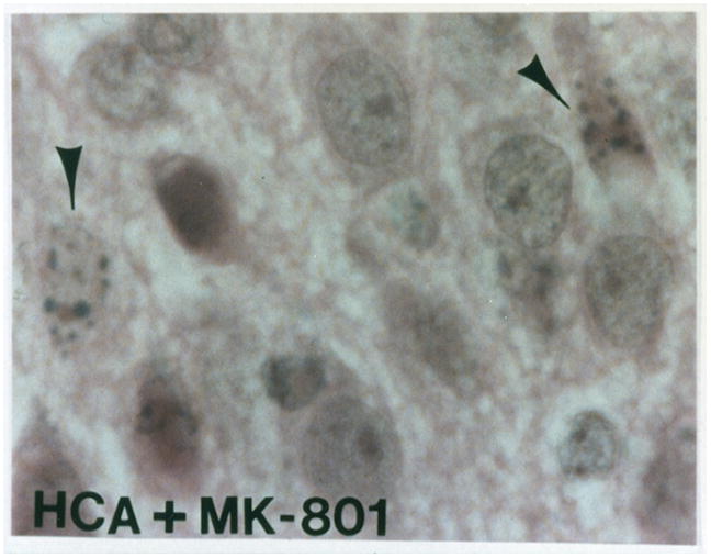
Light microscopy of H&E stained sections for a normal dog (figure 2A). Apoptosis is seen in an HCA treated dog (figure 2B). Reduction in apoptosis seen in an MK-801 treated HCA dog shown by light microscopy of H&E stained sections (figure 2C).