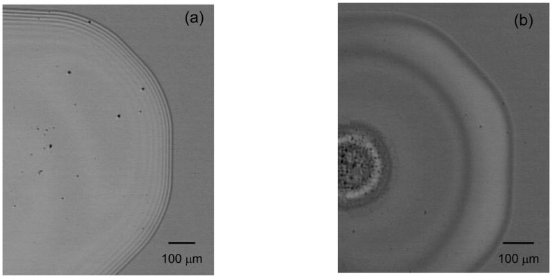
Figure 3.

Microscope images of aqueous HA dried drops at (a) high and (b) low HA concentrations. At high concentrations, concentric rings formed on the outer edge of the drop and the HA dried as a uniform deposition. At low concentrations, HA dried as a ring deposit and no concentric rings were observed. Destructive interference of light resulted in dark bands in microscope images.
