
Figure 4.
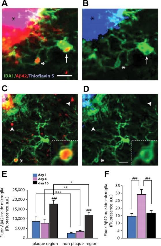
Microglia are effective at phagocytosis of noncongophilic amyloid aggregates in vivo. A, Confocal images of brain slices obtained 4 d after in vivo subarachnoid injection of fluorescently labeled-Aβ42 (red) in CRND8/CX3CR1+/− mice. IBA-1-labeled microglia (green) near amyloid plaques have prominent vesicular structures inside their soma (arrow) and processes (white asterisk) that contain fluorescent Aβ42 (red). Also notice the strong binding of Aβ42 to amyloid plaques (red, black asterisk). B, Thioflavin-S strongly labels the same amyloid plaque (black asterisk). However, despite maximally increasing the blue channel intensity, no thioflavin-S fluorescence can be detected within microglia vesicles that contained Aβ42 fluorescent deposits. C, At 4 d after Aβ42 infusion, most Aβ deposits (white arrowheads) appeared outside microglia, but a substantial number of Fluor-Aβ42 aggregates were already engulfed and found within microglia processes and cell bodies (white asterisk; inset). D, Increasing the blue channel brightness to saturation demonstrates that neither engulfed nor extracellular Fluor-Aβ42 deposits colabeled with thioflavin-S, suggesting that these are newly formed protofibrillar Aβ deposits. Scale bar, 5 μm. E, Quantification of Aβ42 fluorescence (red) shows a greater Aβ42 content within microglia located near the plaque edge (within a 25 μm radius) regardless of the postinfusion interval (***p < 0.001, **p < 0.01, *p < 0.05, two-tailed Student's t test). Sixteen days after infusion, an approximately twofold increase in Aβ42 was observed inside microglia (###p < 0.001, two-tailed Student's t test; n = 39–83 microglia from 3 mice per postinfusion time point; values are expressed as mean ± SEM). F, Fluorescence accumulation outside of microglia undergoes an approximately twofold increase 4 d after infusion but declines again at 16 d (###p < 0.001, two-tailed Student's t test; n = 3 mice per postinfusion time point; values are expressed as mean ± SEM).