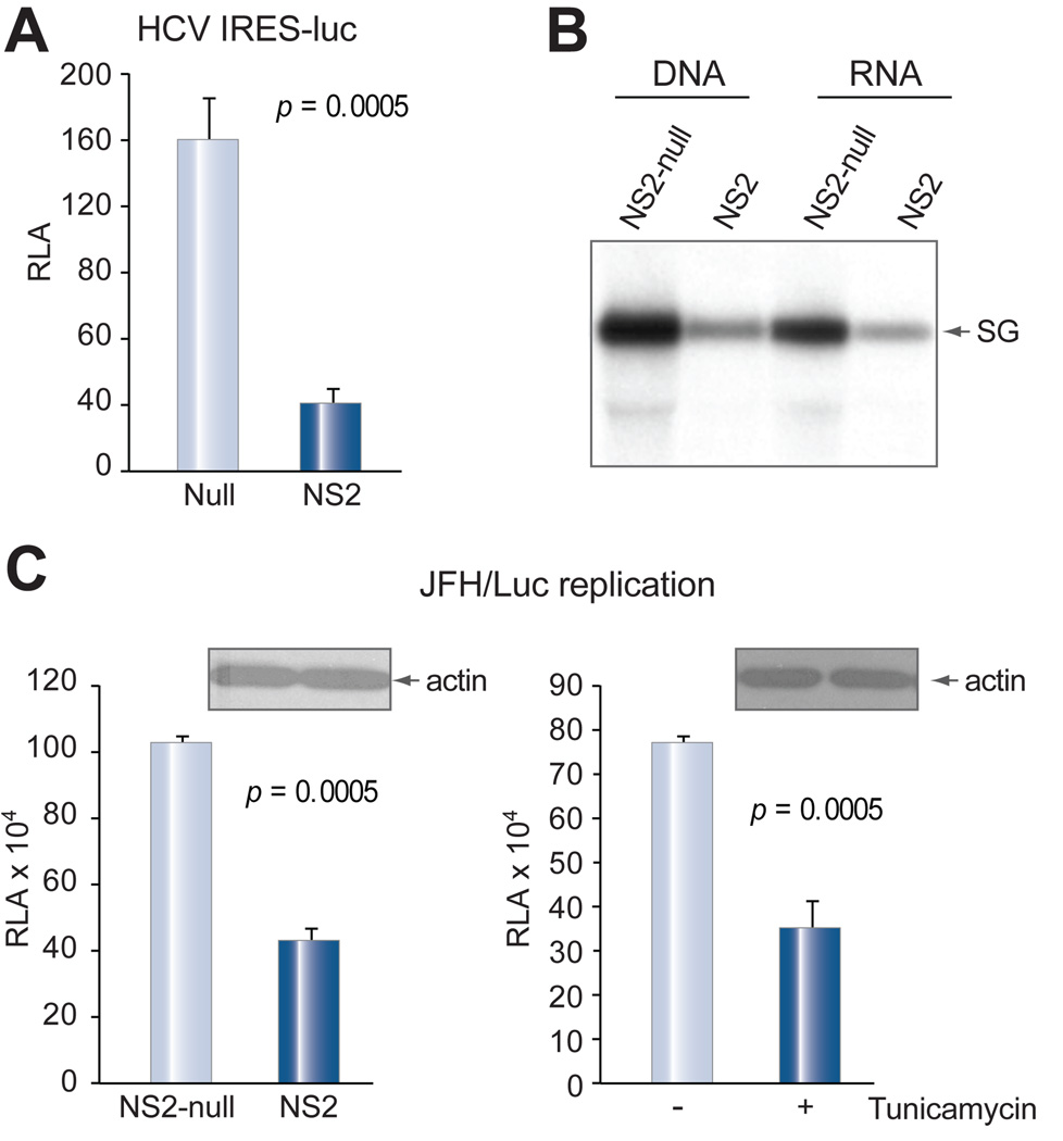
Fig. 8. NS2 protein inhibited IRES-driven protein translation and HCV genome replication.
(A) Protein translation driven by the HCV 5' UTR. Huh-7 cells were electroporated with 5 µg of in vitro transcribed RNA of HCV 5’ UTR-luciferase construct, together with 1 µg of NS2 cDNA or its null mutant. Luciferase was measured 2 days later. Data are presented as mean ± SD (n = 4) and adjusted by transfection efficiency. (B) Replication of susbgenomic replicon (Con1). Huh-7.5 cells (6×10⁶) were electroporated with 1 µg each of subgenomic replicon RNA and NS2 cDNA or its null mutant (left two lanes), or the replicon RNA together with 5 µg of total RNA derived from Huh-7 cells that had been transfected with the corresponding DNA for 2 days (right two lanes). HCV replication was detected 3 days later by Northern blot. (C) Replication of JFH-luc replicon. Huh-7.5 cells (6×10⁶) were electroporated with 5 µg of replicon RNA together with NS2 cDNA or its null mutant (1 µg), and seeded in quadruplicate. Viral replication was measured at day 3 post electroporation by luciferase reporter assay. Alternatively, Huh-7.5 cells were electroporated with replicon RNA alone, and seeded in quadruplicate in two sets. One set was treated with tunicamycin (5 µg/ml) for 24 hrs. Viral replication was measured 3 days later.

