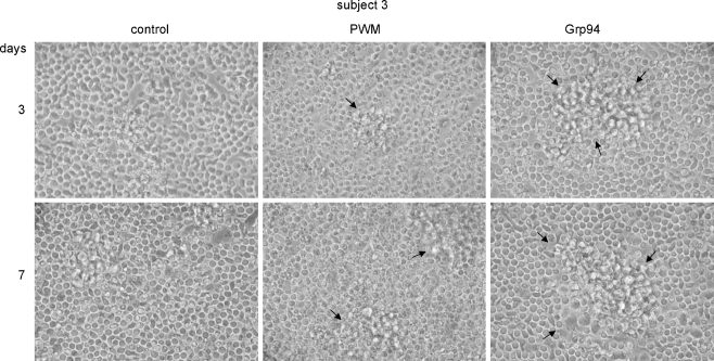
Fig. 5.
Morphological changes of PBMCs in the presence of Grp94. PBMCs were cultured as specified in the “Materials and methods” and at the indicated incubation times morphology was analyzed at the optical microscopy. Representative pictures of many others made on the same well for subjects 3 (as representative of others) are shown in each panel, in the absence (control) and presence of Grp94 (10 ng/ml) and PWM. Big aggregates of large and adherent cells (arrows) characterize PBMCs in the presence of Grp94. More numerous aggregates of similar size were also observed with 100 ng/ml Grp94 (data not shown)

