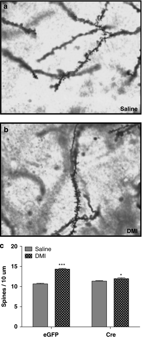
Figure 6.
Hippocampal CA1 dendritic spine density following DMI treatment. (a, b) Representative photomicrographs of Golgi impregnation staining to visualize dendritic arborization following saline (a) or DMI (b) treatment in AAV.eGFP-injected mice. (c) Chronic DMI treatment significantly increased CA1 dendritic spine density in AAV.eGFP-injected mice (***p<0.001, n=30–42 per group). This effect was attenuated in AAV.Cre-injected mice. (*p<0.05). Error bars indicate SEM.

