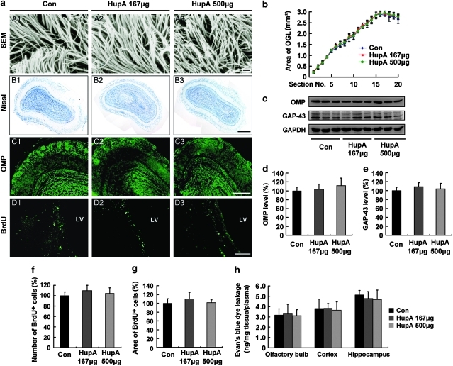
Figure 1.
Evaluation of the potential toxicity of intranasal administration of nasal gel HupA on olfactory bulb (OB) and neurogenesis in the subventricular zone (SVZ) of mice. (a; A1–A3) Scanning electron microscope (SEM) photographs showing no marked changes in the structure of nasal mucocilia between C57BL/6 mice given nasal gel (A1), and nasal gel containing HupA at a dose of 167 μg/kg (A2) and 500 μg/kg (A3), respectively, for 7 days. Scale bar=1 μm. (B1–D3) APP/PS1 mice were treated with vehicle and nasal gel HupA, respectively, for 1 month. Nissl staining of OB exhibited similar histology between vehicle group (B1) and nasal gel HupA at doses of 167 μg/kg (B2) and 500 μg/kg groups (B3). Scale bar=200 μm. (C1–C3) Immunostaining images showing the distribution and expression of olfactory marker protein (OMP) in OB of APP/PS1 mice. No marked changes were found between vehicle- and HupA-treated groups. Scale bar=100 μm. (D1–D3) BrdU immunofluorescent staining showed a similar neurogenesis in SVZ between vehicle and nasal gel HupA groups. Scale bar=50 μm. (b) Measurement of the area of olfactory granule cell layer (OGL) in serial sections of Nissl staining. No significant differences were found between vehicle- and HupA-treated transgenic mice. (c) Western blot showed the protein levels of OMP and GAP-43 in the OB of vehicle- and HupA-treated APP/PS1 mice. GAPDH was used as an internal control. (d, e) Quantification of the protein levels of OMP (d) and GAP-43 (e) in the OB. Intranasal administration of HupA did not alter the expression levels of OMP and GAP-43, compared with controls. (f, g) Quantification of the number (f) and the area (g) of BrdU-labeled cells in SVZ showed no significant difference between control and HupA groups. (h) Leakage of Evan's blue (EB) showed that nasal gel HupA treatment did not significantly change the EB absorbance in the OB, cortex, and hippocampus in APP/PS1 mice. All values are mean±SEM (n=6).

