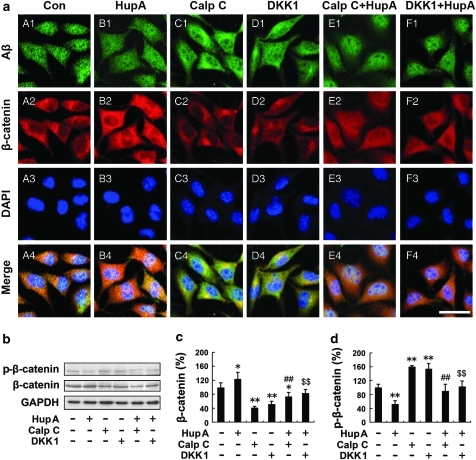
Figure 9.
HupA increases the level of β-catenin in SH-SY5Y cells transfected with APPsw. (a) Double labeling of Aβ (A1–F1) and β-catenin (A2–F2) and confocal microscopic images showing the distribution and expression of β-catenin in APPsw cells. HupA treatment significantly increased the β-catenin immunofluorescence in APPsw cells (B2), compared with the control (A2). Calphostin C or DKK-1 treatment decreased the levels of β-catenin (C2 and D2). Additional HupA treatment reversed the reduction of β-catenin in cultures pretreated with inhibitors Calphostin C or DKK-1 (E2 and F2). Scale bar=30 μm. (b) Western blot analysis showing the levels of β-catenin and p-β-catenin in APPsw cells treated with HupA or inhibitors. GAPDH was used as an internal control. (c, d) The protein level of total β-catenin was significantly increased, whereas the level of p-β-catenin was significantly decreased in HupA-treated cells compared with controls. There was a significantly reduced level of total β-catenin and an increased level of p-β-catenin in cultures treated with Calphostin C or DKK1, whereas the total β-catenin level was increased and the p-β-catenin level was reduced when cultures were preincubated with inhibitors followed by HupA. Representative immunoblots from three experiments are shown. The data shown in (c, d) are presented as mean±SEM of at least three independent experiments. **p<0.05, **p<0.01 vs control group; ##p<0.01 vs Calphostin C-treated group; $$p<0.01 vs DKK1-treated group.

