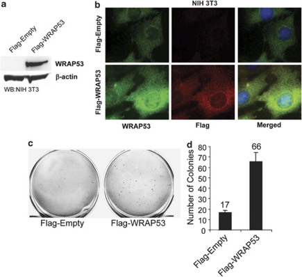
Figure 2.
WRAP53 overexpression leads to cell transformation. (a) WB analysis of WRAP53 protein levels in NIH 3T3 cells transfected with the indicated vectors. (b) IF of NIH 3T3 cells using WRAP53 and Flag-specific antibodies. Cells were transfected with the indicated plasmids and selected in the presence of 1 μg/ml G418 for 9 days before staining. Nuclei were stained with DAPI in all IF stainings. (c) Anchorage-independent transformation assay of NIH 3T3 cells expressing Flag-tagged WRAP53 or Flag-Empty. The cells were transfected with the indicated plasmids and selected in the presence of 1 μg/ml G418 for 9 days. Selected cells were suspended in soft agar and were photographed after 19–23 days incubation at 37°C. (d) The graph shows the mean of two independent experiments; error bars represent standard error

