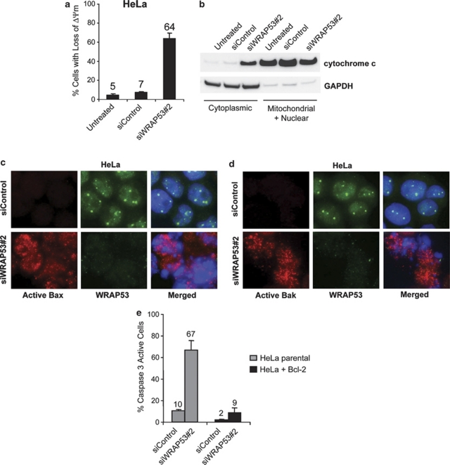
Figure 4.
WRAP53 knockdown induces mitochondrial-dependent apoptosis. (a) The bars show the percentage of TMRE-negative HeLa cells treated with siControl or siWRAP53 no.;2 for 72 h. Error bars represent standard error of three independent experiments. (b) WB analysis of cytochome c and GAPDH levels in cytoplasmic and mitochondrial/nuclear fractions of HeLa cells treated with the indicated siRNA oligos for 72 h. GAPDH was used as a cytoplasmic marker. (c and d) IF staining of WRAP53 and active Bax (c) or WRAP53 and active Bak (d) in HeLa cells treated with siControl and siWRAP53 no.;2 oligos for 72 h. Cytospin preparations have been used for Bax and Bak IF experiments resulting in a skewed subcellular distribution of WRAP53. Normally, the majority of WRAP53 protein is concentrated in the cytoplasm of HeLa cells similar to the staining pattern in NIH 3T3 cells (Figure 2b) (e) The bars show the percentage of active caspase 3-positive parental HeLa or Bcl-2 overexpressing (HeLa+Bcl-2) HeLa cells, treated with siControl and siWRAP53 no.;2 for 72 h. Error bars represent standard error of three independent experiments

