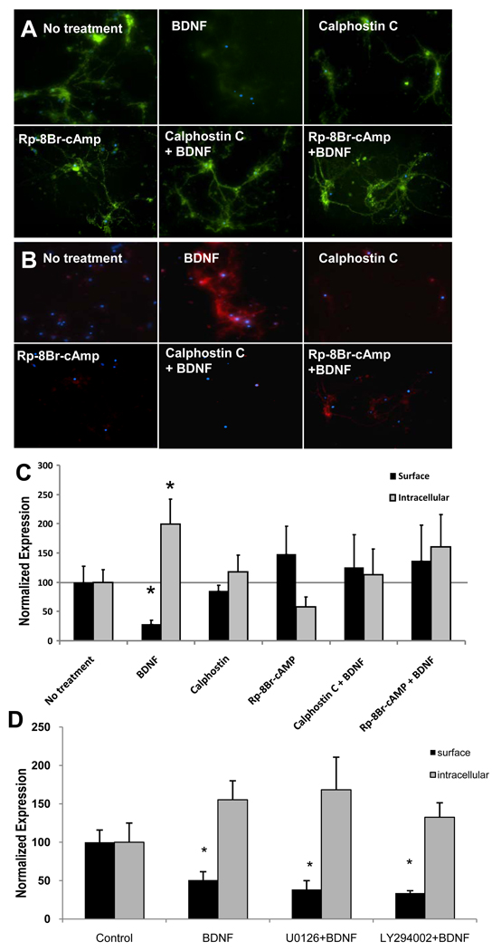
Figure 4. BDNF-dependent decrease in surface GABAARα1 subunits is reversed by treatment with PKA and PKC inhibitors.
Panel A illustrates light microscopic images of surface (green) GABAARα1 subunits following treatment with vehicle, BDNF, Calphostin C (PKC inhibitor), Rp-8Br-cAMP (PKA inhibitor), or these agents combined with BDNF. Panel B illustrates images of internalized (red) GABAARα1 subunits following the same conditions as in (A). Panel C demonstrates quantification of fluorescence intensity of surface (black) or intracellular (gray) GABAARα1, normalized with controls. Panel D demonstrates quantification of fluorescence intensity of surface (black) or intracellular (gray) GABAARα1, normalized with controls following inhibitors of MAPK (U0126) or PI3K (LY294002), neither of which had an effect on BDNF-induced GABAARα1 internalization. (Mean ± SEM of normalized intensity, * p<0.05, relative to no treatment).

