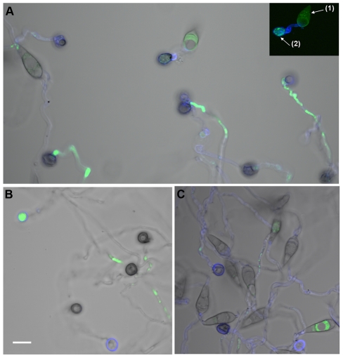
Figure 7. Δhyr1 (B25) conidia on gel-bond were similar to wild type in terms of ROS production.
Staining was performed 24 hpi; Calcofluor White was used to stain the cell walls (blue) and H2DCFDA was used to stain the ROS (green). Conidia of (A) Δhyr1 (B25), (B) wild type (70-15) and (C) ectopic (B40). A transmitted light image was taken as well, and overlaid with the fluorescent image. The inset in panel A showed the fluorescence image of the conidium (1) and appressorium (2). Images were taken using confocal microscopy. Scale bar = 10 µm.

