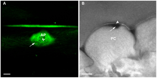
Figure 8. Δhyr1 appressorial-localized ROS appeared to be plant-generated.
(A) Reflection confocal imaging with the ROS stain DAB shows a wide ROS signal (arrow) around and beneath the appressorial attachment site (AP). In the middle of the appressorium attachment site was the putative penetration peg site (arrowhead). (B) The same interaction site as Fig. 8A, embedded in epoxy resin and imaged under confocal microscopy revealed DAB deposited (arrow) beneath and surrounding an attempted penetration site (arrowhead). The deposit was located up against the plant cell wall (PC) on the inside of the cell. Scale bar = 5 µm.

