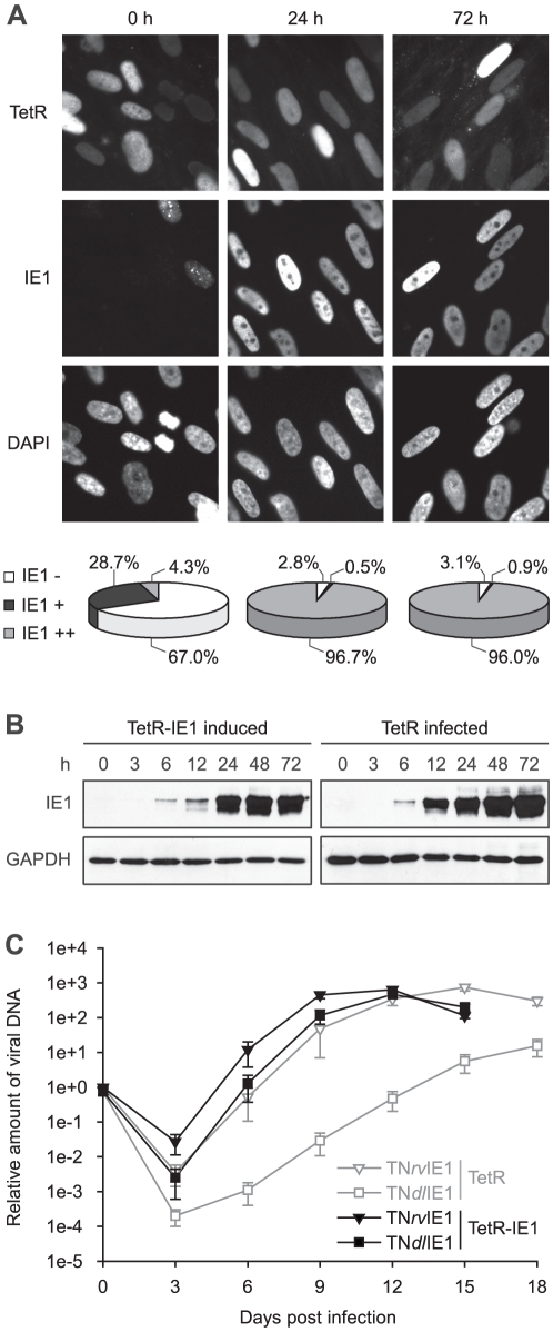
Figure 1. Characterization of TetR-IE1 cells.
A) TetR-IE1 cells were treated with doxycycline for 24 and 72 h or were left untreated (0 h). Paraformaldehyde-fixed samples were examined by fluorescence microscopy for IE1 (antibody 1B12) and TetRnlsEGFP (TetR) expression (autofluorescence). Staining with 4′,6-diamidino-2-phenylindole (DAPI) was performed to visualize nuclei. Original magnification, ×504. For the pie charts, ∼500 randomly selected nuclei per sample were examined for IE1 expression. The scoring system is as follows: IE1 −, no IE1 staining above background; IE1 +, weak, mostly punctate IE1 staining; IE1 ++, strong, diffuse IE1 staining. B) Time course (0–72 h) immunoblot analysis of IE1 and GAPDH steady-state protein levels in doxycycline-induced TetR-IE1 cells and hCMV (TNwt)-infected TetR cells (MOI = 1 PFU/cell). To assure comparability between protein bands, gels loaded with extracts from equal cell numbers were run and blotted side by side under the same conditions, and pairs of membranes destined for IE1 or GAPDH detection were processed together and exposed on the same film. C) Multistep replication analysis of IE1-null mutant hCMV (TNdlIE1) and the corresponding revertant virus (TNrvIE1) in doxycycline-treated TetR and TetR-IE1 cells. Confluent cells were infected at an MOI of 0.01 PFU/cell, and viral replication was monitored at 3-day intervals by qPCR-based relative quantification of hCMV DNA from culture supernatants. Mean values and standard deviations of four independent infections with two different clones per each virus strain are shown.

