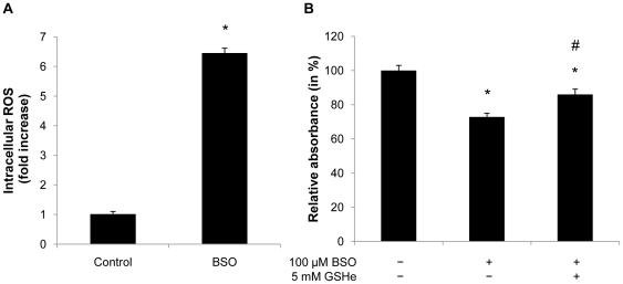
Figure 3. Glutathione depletion induces intracellular ROS formation during differentiation.
A: Confluent 3T3-L1 cells were treated with 100 µM BSO. After 2 days BSO treatment was renewed and cells were induced to differentiate. 24 h after hormonal induction cells were incubated with 10 µM H2DCFDA and analyzed by FACS. B: Confluent 3T3-L1 cells were treated with 100 µM BSO until day 2 of differentiation. 5 mM GSH-ester was added one day prior to differentiation. On day 7 of differentiation cells were stained with Oil-red-O and analyzed using a spectrophotometer. All results are presented as mean ± SEM (* p<0.05 vs. control, # p<0.05 vs. BSO).

