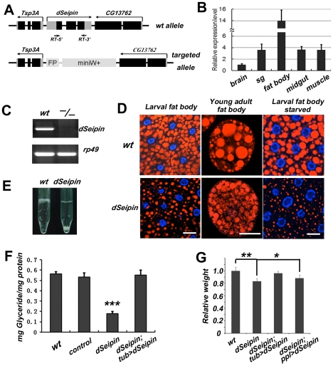
Figure 1. dSeipin mutants exhibit reduced lipid storage in the fat body.
(A) Schematic of the genomic structures of wild type dSeipin and the null mutant. In the null mutant, the dSeipin locus is replaced by GFP (FP) and miniWhite (miniW+) sequences. Except for the FP and miniW+ regions, black boxes represent coding regions and grey boxes represent un-translated region. (B) Expression levels of dSeipin in different larval tissues by qRT-PCR. The error bars represent standard deviation. (C) RT-PCR analysis of wild-type and dSeipin knockout larvae. Primer positions (RT-5′ and 3′) are labeled in (A). (D) Lipid droplets labeled by Nile red (red) in larval fat bodies and young adult fat cells from wild type and dSeipin mutants. Nuclei were stained with DAPI (blue). dSeipin mutants exhibit lipid storage defects with small lipid droplets. Under starved conditions the lipid droplets in dSeipin mutant larvae are even smaller. Scale bars: 20 µm. (E) Larval fat bodies from wild type float on top of 2% sucrose solution, while fat bodies from dSeipin mutants sink to the bottom. (F) Glyceride levels in adult males of wild type, control, dSeipin mutants and transgene-rescued dSeipin mutants. dSeipin mutants have significantly lower levels of glyceride. The error bars represent standard deviation. ***: P<0.0001. (G) Average weights (adult male) from wild type, dSeipin mutants and transgene-rescued dSeipin mutants. dSeipin mutants are slightly lower in weight. The error bars represent standard deviation. **: P<0.001; *: P<0.05.

