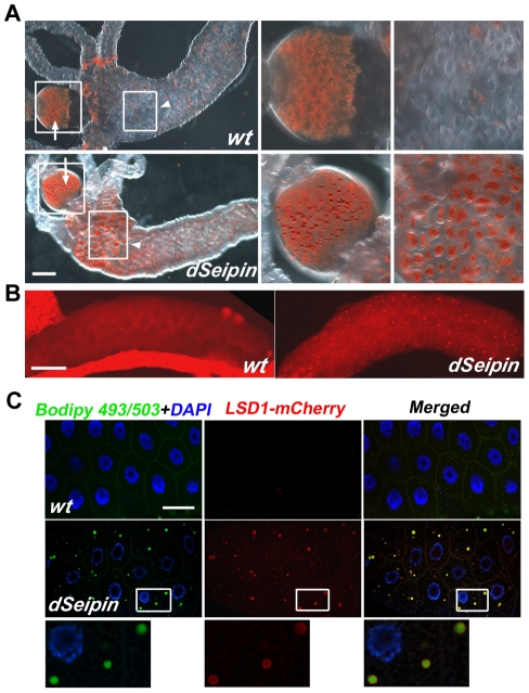
Figure 3. Ectopic lipid droplet formation in dSeipin mutants.
(A) Oil Red O staining in guts of wild type and dSeipin mutants. In wild type the proventriculus (arrow) has numerous small droplets and the anterior midgut (arrowhead) has only a few Oil Red O-positive patches. In dSeipin mutants, the droplets in the proventriculus (arrow) are larger than wild type and in the anterior midgut (arrowhead) there are more patches of Oil Red O-positive staining. The boxed regions of the proventriculus and the midgut are enlarged in the adjacent panels. Scale bar: 100 µm. (B) Nile red staining in salivary glands. In wild-type salivary glands, essentially no punctate Nile red staining was found. In dSeipin mutants, many Nile red-positive puncta are present, indicating ectopic lipid storage. Scale bar: 100 µm. (C) Ectopic lipid puncta marked by Bodipy 493/503 (green) in dSeipin mutant salivary glands are confirmed as lipid droplets by co-labeling with the lipid droplet surface marker LSD-1-mCherry (red). Nuclei were stained with DAPI (blue). Scale bar: 50 µm. The lower panels show enlarged views from a dSeipin mutant. Expression of LSD-1-mCherry in a wild-type background was used as a control.

