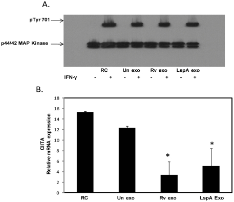
Figure 4. Exosomes from M.tb-infected cells do not block IFN-γ induced STAT1 phosphorylation but do inhibit IFN-γ induced expression of CIITA.
BMMØ were treated +/− exosomes isolated from RAW264.7 macrophagesas described for figure 1 followed by a 30 minute incubation with IFN-γ. Cells were lysed and analyzed by Western blot for p-STAT1 (Tyr701) (A). The p44/42 MAP Kinase antibody was used as a loading control as described previously (17). BMMØ were treated with exosomes and stimulated +/− IFN-γ for 18 hours. Cells were harvested for qRT-PCR using specific primers for target gene (CIITA) and reference gene (GAPDH). Shown is the relative mRNA expression compared to untreated cells for CIITA normalized to GAPDH (B). Results are representative of two separate experiments plus standard deviation and p value <0.05 shown by asterisk (*).

