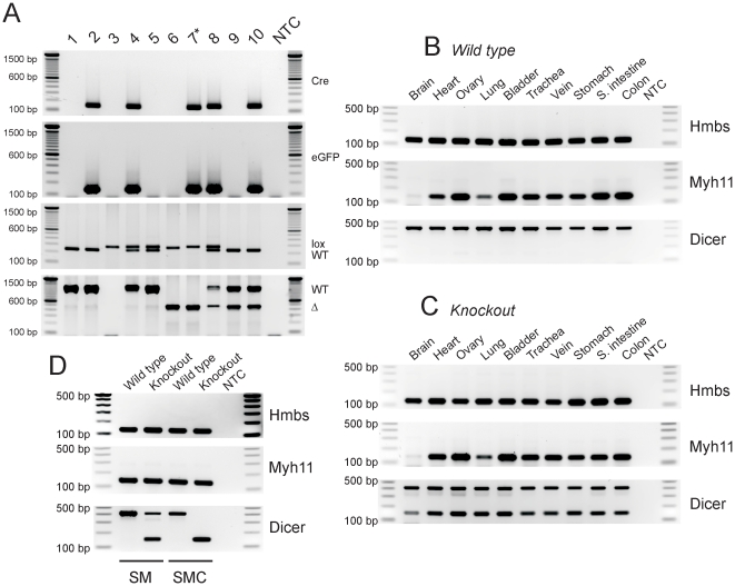
Figure 1. Genotyping and expression analysis of SMC-specific Dicer knockout mice.
(A) Each mouse line was genotyped using PCR with a set of primers specific for Cre, eGFP, inserted lox P sites (lox), and wild type (WT) or deleted locus (Δ). The smDicer homozygous knockout (KO) smDicer−/−;Cre-GFP/+ is indicated by an asterisk on the mouse line 7 (★). All genotypes for mouse lines 1–10 are shown in Table 1. (B) Myh11 and Dicer transcripts abundantly expressed in the SM organ tissues from the WT mouse. Hmbs was used as an endogenous control. (C) Expression of Myh11 and Myh11-dependent KO (deletion) of Dicer transcripts in the SM organ tissues from the smDicer KO mouse. Note that the deleted Dicer PCR products (∼150 bp) are smaller than the WT (∼400 bp). (D) Expression of Myh11 and Dicer transcripts in the small intestine SM tissue (SM) and cells (SMCs) sorted by FACS. Dicer transcripts were completely deleted in the sorted SMCs from the smDicer KO mouse while they were partially deleted in the SM. All PCR products were analyzed on 1.5% agarose gels along with a DNA ladder. NTC stands for non-template control, SM for smooth muscle tissue, and SMC for smooth muscle cells.

