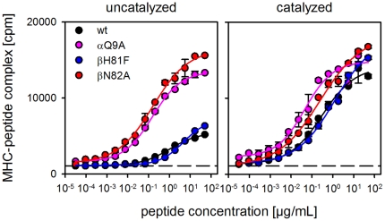
Figure 5. Influence of αQ9 and βN82 on the receptiveness of soluble HLA-DR1.
ELISA-based antigen loading assays were carried out with soluble forms of wt HLA-DR1 (black) and mutants αQ9A (magenta), βN82A (red), and βH81F (blue). MHC molecules derived from insect cells were loaded with biotinylated peptide HA306-318. Loading was carried out in the absence (left panel) or presence (right panel) of the ‘MHC-loading enhancer’ (MLE) compound AdEtOH [10]. The dashed line indicates the background in the absence of ligand.

