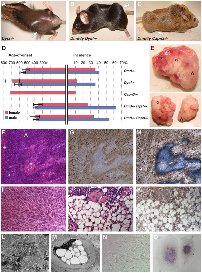
Figure 1. MD mice are prone to develop skeletal muscle-related malignant mixed mesenchymal tumors.
(A) Dysferlin-deficient mouse (male, 878 d) with sarcoma located in the caudal abdominal wall (arrow; weight of excised tumor: 4.2 g). (B) Dystrophin-dysferlin double-mutant (male, 250 d) with very large sarcoma (9.1 g), which arose in proximal and distal skeletal muscles of the left hindlimb (arrow). (C) Dystrophin-calpain 3 double-mutant (male, 474 d) with sarcoma (2.3 g) located in ischiocrural muscles of the right hindlimb (arrow). (D) Graphical representation of sarcoma incidence and age-of-onset from life-span studies of mice lacking dystrophin (n = 48/122; C57BL/10), dysferlin (n = 35/151; C57BL/10), or calpain 3 (n = 1/19; 129/Sv x C57BL/6) and of dystrophin-dysferlin (n = 42/89) and dystrophin-calpain 3 (n = 24/55) double-mutant mice. Error bars indicate confidence interval of the mean (95%). (E) Representative examples of excised tumors (from dystrophin-dysferlin double-mutant mice) with myo- (*), lipo- (°), and fibrosarcomatous (^) compartments macroscopically recognizable as different parts of the tumor mass. (F–H) Histological examination of the tumor shown in E (upper panel) revealed a sarcoma with mixed mesenchymal differentiation characterized by rhabdomyo- (*), lipo-(°), and fibrosarcoma (^) components. Serial sections used for H&E staining (F), myogenin staining (G), and double staining with myogenin and Sudan Black (H). (I) Morphological analysis of a sarcoma that arose in a 486 d-old C57BL/10 mdx mouse revealing a prominent fibrosarcoma areal characterized by the typical collagen fibres bundles arranged in “herringbone pattern” (H&E). (J,K) Well-differentiated liposarcoma area within a mixed sarcoma from a Dmd −/− Capn3 −/− double-mutant mouse (J, H&E), with intense Cdk4-positivity (K). (L) Electron micrograph showing a rhabdomyosarcoma cell displaying myofibril structures. (M) Electron micrograph from the same tumor showing a liposarcoma cell densely packed with large lipid vesicles and exhibiting a highly aberrantly shaped nucleus. (N) Photomicrograph showing cultured tumor cells propagated from a typical mixed sarcoma (from a dysferlin-deficient mouse), indicating the presence of different types of tumor cells (round-shaped, spindle cell-like, and myotube-like elongated rhabdomyoblasts). (O) Photograph of two tumor cells grown in vitro from the same explanted tumor. Double-staining for myogenin and Sudan Black depicted a myogenin-positive myogenic tumor cell next to a myogenin-negative but lipogenic tumor cell containing Sudan Black-positive droplets.

