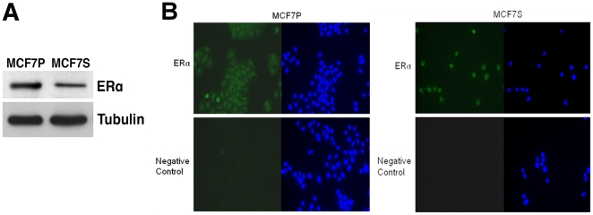
Figure 3. Estrogen receptor status in MCF7S.
A. Immunoblotting of ERα protein in MCF7P and MCF7S cells. Tubulin served as loading control. B. Indirect immunofluorescence for ERα protein (green) in MCF7P and MCF7S. Cells were fixed by 3.7% formaldehyde. HOECHST 33342 (blue) was used to indicate nuclear region. A negative control was performed without primary anti-ERα antibody. Magnification at 40x.

