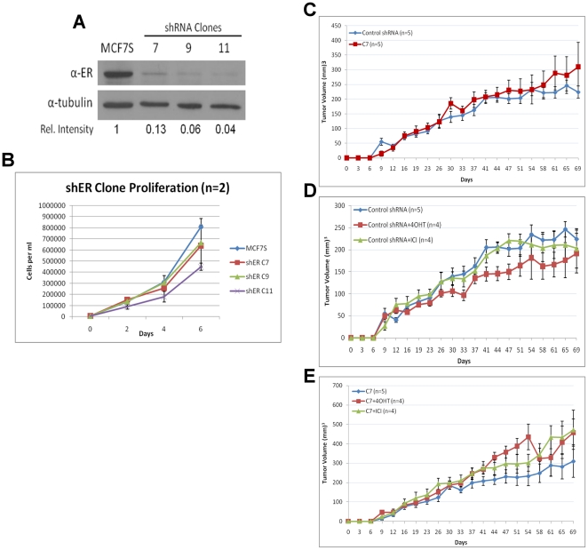
Figure 8. ERα is disposable in MCF7S.
A. Immunoblot analysis of ERα in three shERα MCF7S clones. Relative densitometry intensity is shown. B. Cell proliferation of three shERα clones compared to bulk MCF7S culture. Error bars represent standard deviation from two experiments. C. In vivo tumorigenic assay comparing shRNA control and shERα knockdown MCF7S. Five mice were injected for each condition. D and E. In vivo tumorigenic assay comparing antiestrogen treated (4 days) shRNA control or shERα MCF7S cells. At least 4 mice were injected for each condition.

