Fig. 1.
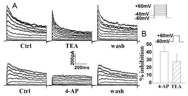
Expression of outward K+ current in rat microglia. Panel A shows examples illustrating the voltage-dependent outward K+ current recorded in rat microglia and the partial blockade of outward K+ current by TEA (upper) and 4-AP (lower). Panel B depicts the average inhibition of whole-cell outward K+ current in microglia by 4-AP and TEA when measured at command voltage step of +60mV. Bars represent mean±SEM (the same in the following figures unless indicated). Voltage protocol employed to generate outward K+ current is shown above Panel B.

