
Figure 3. The proteostasis boundary (PB).
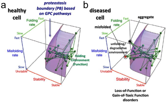
The position of each node (protein) relative to the PB (curved surface) responsible for biological function is defined by a protein’s folding properties: folding kinetics (Z-axis), misfolding kinetics (Y-axis) and thermodynamic stability (X-axis). Each line defines a physical or functional interaction between two proteins in the system. The location of the PB in 3D space is established by the composition of the PN and modulated by the GPC triad. (a) All of the nodes are within the PB boundary in a healthy cell. (b) Mutations or aberrant post-translational modifications can alter folding kinetics and energetics, making their corresponding nodes and edges fall outside (above the curved surface) of the PB. This space in the 3D plot does not support function of the energetically destabilized variant and can lead to either degradation (red node) or protein aggregation (black node). The loss of connectivity to proteins within the embrace of the PB can challenge the entire PN leading to cell, tissue, and organismal disease. Reproduced with permission from Elsevier Press [2].