
Figure 1.
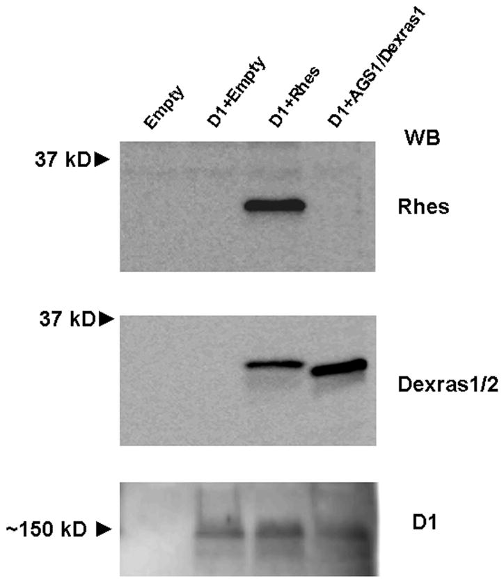
Expression of Rhes, AGS1/Dexras1, and D1 receptors in CHO cells. Shown are Western blots of cell lysates (20 μg total protein) of CHO cells assayed 24 hours after transfection with either empty vector, or a combination of D1 and either Rhes or AGS1/Dexras1. Empty vector-transfected cells did not express any of the proteins. A ~30 kD protein was detected by the anti-Rhes antibody in Rhes-transfected cells, whereas the antibody to Dexras1/2 detected a ~30 kD protein in cells transfected with either Rhes or AGS1/Dexras1. D1 receptor was detected in all D1-transfected cells, in a large, likely oligomeric, form. Blots shown are representative of 4 assays. WB = Western blot.