Fig. 7.

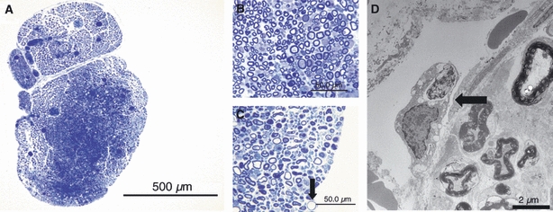
At 2 days after removal of the epi-perineurium, marked endoneurial swelling in the outer portion was observed in sections stained for toluidine blue (bar = 500 μm) (A). In the central portion of the nerve fascicle, most of the myelinated nerve fibers remained intact (bar = 50 μm) (B). However, in the swollen outer portion, degeneration of the nerve fibers was noted (arrow) (bar = 50 μm) (C). An englobed myelinated nerve (arrow) outside the endoneurium was observed by electron microscopic examination (bar = 2 μm) (D).
