
Figure 1.
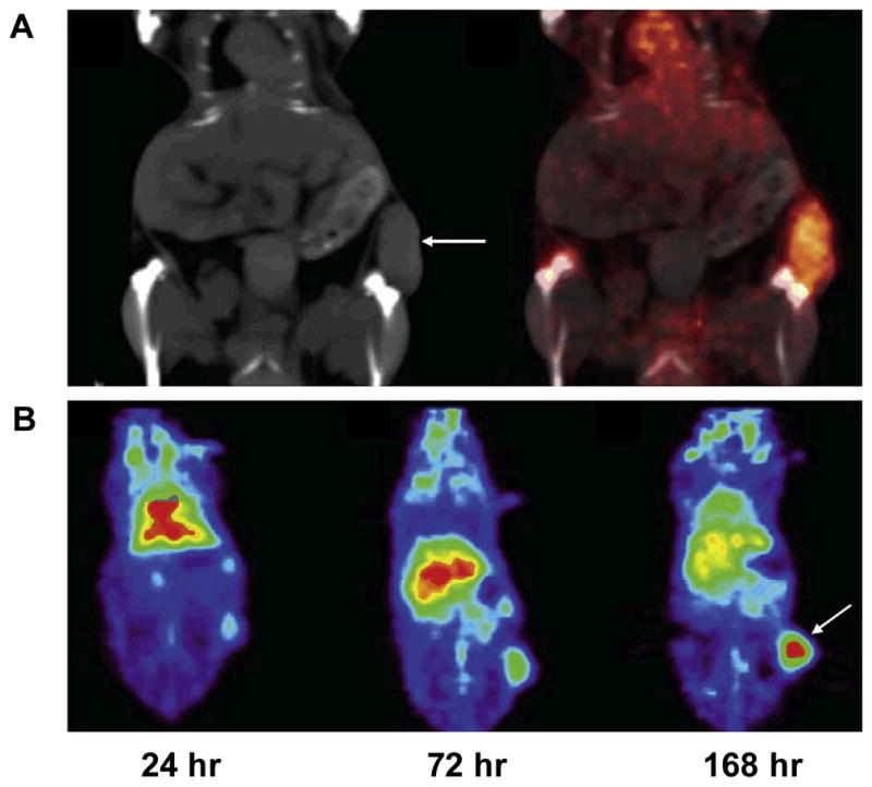
Noninvasive imaging of VEGF expression in vivo by 89Zr-labeled VEGF antibody Bevacizumab. A. Coronal CT image (left) with clear subcutaneous localization of SKOV-3 tumor (arrow) and fusion of microPET and CT images (right) (168 h after injection) (arrows indicate SKOV- 3 tumors). B. Coronal planes of microPET images at different time points after 89Zr-Bevacizumab injection. Reproduced from reference 52 with permission.