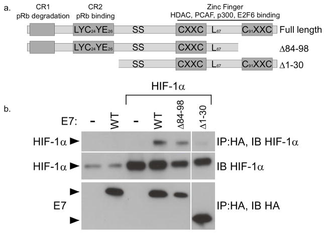
Figure 3. Mapping the HIF-1α binding domain of E7.
(a) A schematic of the E7 protein, showing important functional domains and amino acids that were mutated in this study. (b) U2OS cells were cotransfected with the indicated 16E7 deletion construct and an expression vector for HIF-1α. Following 24 hours incubation, cells were treated with 100 μM DFO for 16 hours and total lysates prepared. E7-containing complexes were immunoprecipitated with anti-HA antibodies. HIF-1α was detected by SDS/PAGE and western blotting of the immunoprecipitates.

