
FIGURE 1.
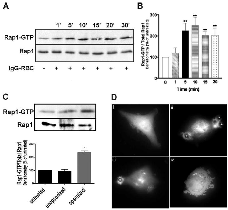
FcγR ligation activates Rap1. NR8383 macrophages were incubated with 30/1 (A) IgG-opsonized or (C) unopsonized SRBCs for varying time points, lysed, and assayed for GTP-bound Rap1 as described in Materials and Methods. Total levels of Rap1 in cell lysates are shown (bottom blot). No crossreactivity was observed between the Rap1 Ab and SRBC proteins (data not shown). B, Densitometric analysis of Rap1 pulldowns from cells challenged with IgG-opsonized SRBCs was performed as described in Materials and Methods. Results represent the means (±SE) for 10 independent experiments. **, p < 0.01 compared with control by ANOVA followed by Dunnett’s multiple comparison test. D, GFP-RalGDS-transfected macrophages were overlaid with 10/1 IgG-opsonized SRBCs (denoted by asterisks) and imaged using fluorescence microscopy at (i) 0 min, (ii) 5 min, and (iii) 10 min. Negative control GFP-transfected macrophages were overlaid with IgG-opsonized latex beads and imaged at (iv) 10 min. Images are representative of at least three separate experiments.