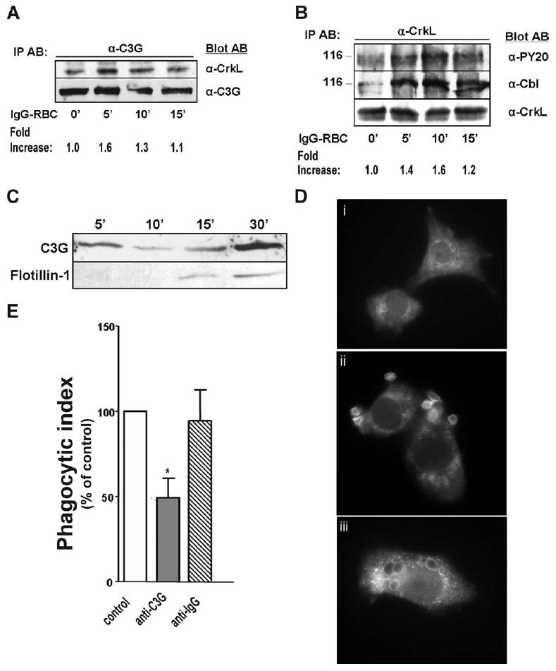
FIGURE 4.

C3G is activated by FcγR ligation and translocates to the phagosome. NR8383 macrophages were incubated with 30/1 IgG-opsonized SRBCs for varying time points, lysed, and immunoprecipitated with (A) anti-C3G and (B) anti-CrkL Abs. Bound proteins were immunoblotted with Abs indicated. Fold increase in (A) CrkL or (B) phosphorylated tyrosine at 116 kDa was measured by densitometric analysis for each of the three Western blots and normalized to unstimulated cells. One blot representative of a total of three is shown. C, Phagosomal membranes were harvested from NR8383 macrophages stimulated with 30/1 IgG-opsonized magnetic beads as described in Materials and Methods. As a late phagosome marker, membranes were probed for flotillin-1 (1/1000, BD Biosciences). D, NR8383 macrophages were overlaid with 10/1 IgG-opsonized SRBCs for (i) 0 min, (ii) 3 min, and (iii) 15 min and immunostained for C3G. Images are representative of at least three separate experiments. E, NR8383 cells incubated with liposome constructs containing anti-C3G or isotype control Ab as detailed in Materials and Methods were assessed for phagocytic ability. Data shown are the means ± SE of three independent experiments. *, p < 0.05 compared with vehicle macrophages by Student’s t test.
