
FIGURE 5.
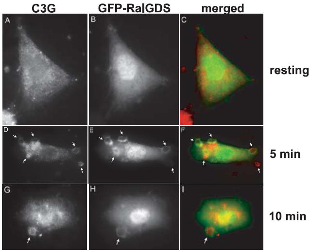
C3G and Rap1GTP both translocate to the phagosome upon FcγR ligation. GFP-RalGDS-transfected macrophages were overlaid with 10/1 IgG-opsonized SRBCs for (B) 0 min, (E) 5 min, and (H) 10 min and immunostained for C3G (A, D, and G). Merged images (C, F, and I) show GFP-RalGDS in green and C3G in red. Arrows indicate location of bound SRBCs on NR8383 cells. Images are representative of at least three separate experiments.