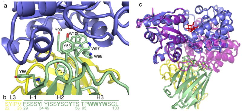
Figure 5. The “wedge” formed by the CDR loops of MCS2.
(A) A group of bulky side-chain residues within CDRH-3 of the sAB (green sticks) form the wedge structure, which interacts with a region within MBP (blue) that is only exposed in the closed, maltose-bound conformation. (B) Sequence of the CDR loops of MCS2. Bold letters indicate residues that interact with the MBP molecule. (C) An overlay of the MCS2-MBP complex with the open form of MBP (purple, PDB code: 1OMP 12) indicates that the apo form of MBP clashes with the sAB CDR loops.

