Figure 5.
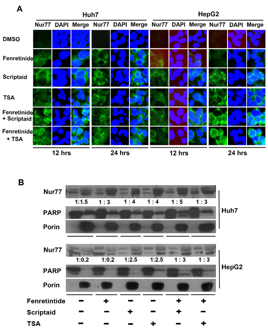
HDACi and/or fenretinide induced cytoplasmic Nur77 expression in HCC cells. (A) HCC cells were treated as described in Figure legend 1. Immunofluorescence staining was performed using anti-Nur77 antibody and nuclear counterstaining with DAPI and viewed by confocal microscopy. (B) Nuclear (Nu) and Mitochondria (Mit) enriched fractions were isolated from treated cells. Proteins were fractionated followed by western blot using antibodies specific to Nur77, PARP, and Porin. Numbers indicate the ratios of nuclear protein vs. mitochondria protein.

