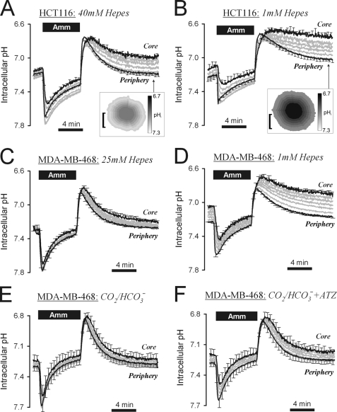
FIGURE 4.
Extracellular mobile buffers facilitate membrane H+ transport in HCT116 and MDA-MB-468 spheroids. A, 20 mm ammonium (Amm) prepulse performed on HCT116 spheroid (mean spheroid radius = 144.9 ± 8.7 μm), superfused with 40 mm Hepes-buffered solution at pH = 7.4. Inset: pHi map (bar = 100 μm). End point pHi gradient = 0.243 ± 0.044. B, experiment repeated with 1 mm Hepes-buffered solution at pH = 7.4 (mean radius 127.7 ± 3.9 μm). Inset: pHi map (bar = 100 μm). End point pHi gradient = 0.314 ± 0.079. C, ammonium prepulse performed on MDA-MB-468 spheroid (mean radius 127 ± 9.8 μm), superfused with 25 mm Hepes-buffered solution at pH = 7.4. End point pHi gradient = 0.118 ± 0.008. D, experiment repeated in 1 mm Hepes-buffered solution. End point pHi gradient = 0.284 ± 0.019. E, experiment repeated with superfusate buffered with 5% CO2/22 mm HCO3− (mean radius 138 ± 13.0 μm). End point pHi gradient = 0.051 ± 0.038. F, experiment continued in 100 μm acetazolamide (ATZ). End point pHi gradient = 0.076 ± 0.039. Significant increase in pHi at core (p = 0.0052) and periphery (p = 0.0315). Error bars in all panels indicate S.E.

