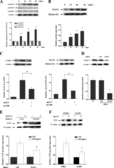
FIGURE 4.
STAT3 activation is involved in the apoA-I-mediated increase of TTP expression in LPS-treated macrophages. A, THP-1 macrophages were treated with apoA-I for different times as indicated. Proteins were extracted, and the phosphorylated STAT1, STAT3, and STAT6 levels were measured by immunoblot analyses with antibodies specific for phosphorylated signal transducer and activator of transcriptions. Values were normalized against β-actin. *, p < 0.05 versus 0 min. B, THP-1 macrophages were treated with apoA-I for different times as indicated. The nuclear proteins extracted from cells were subjected to immunoblot analyses with antibodies against STAT3 and histone H1. *, p < 0.05 versus 0 min. C, THP-1 macrophages were pretreated with apoA-I for 30 min. Thereafter, the medium was replaced with fresh medium containing the AG-490 (30 μm). Then cells were incubated for another 30 min, and total or nuclear proteins were subjected to immunoblot analyses with antibody against p-JAK2 and STAT3. *, p < 0.05. D, THP-1 macrophages were transfected with control (WT) or STAT3 siRNA for 48 h, and protein samples were immunoblotted with STAT3 and β-actin antibodies. *, p < 0.05 versus control group. E, THP-1 macrophages transfected with control or STAT3 siRNA were incubated with LPS for 4 h with or without pretreatment of apoA-I. Protein samples were immunoblotted with TTP and β-actin antibodies. *, p < 0.05 versus control group. F, THP-1 macrophages were pretreated with AG-490 or DMSO for 1 h, and cells were then incubated with LPS for another 4 h with or without pretreatment of apoA-I. Protein samples were immunoblotted with TTP and β-actin antibodies. *, p < 0.05 compared with DMSO group. All of the results are the mean ± S.D. of quadruplicate values from three separate experiments.

