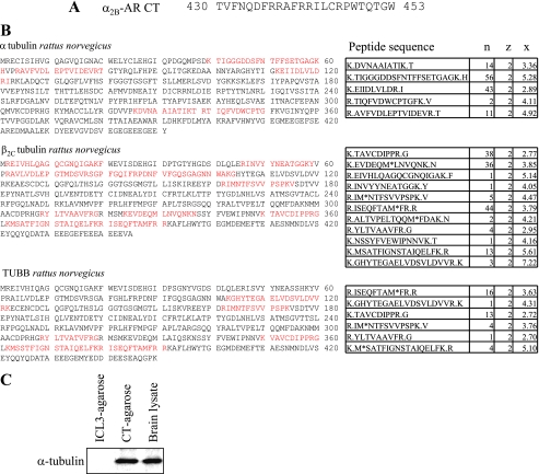
FIGURE 1.
Identification of tubulin isoforms as interacting proteins of the α2B-AR CT. A, sequence of the α2B-AR CT that was directly fused to agarose beads to generate affinity matrix. B, identification of tubulin interacting with the α2B-AR CT-conjugated agarose. CT-conjugated agarose beads were incubated with rat brain lysates and bound proteins were eluted and separated by two-dimensional gel electrophoresis. Spots of interest were picked, digested, and identified by LTQ electrospray mass spectroscopy as described under “Experimental Procedures.” n, number of times the sequence showed up in the spot of interest; z, charge of the peptide sequence; x, correlation score (representation of how well the mass spectrum matches a pre-generated standard for that particular sequence). Peptides are generally considered significant with a correlation score of over 2.5 at a charge of 2, or with a correlation score of over 3 at a charge of 3. Red indicates the positions of the peptides identified by mass spectroscopy in the full length of tubulin isoforms. C, detection of α-tubulin in the eluate from CT- and ICL3-agarose beads. A small portion of the eluate was analyzed by Western blotting using α-tubulin antibodies. Similar results were obtained in at least three separate experiments.

