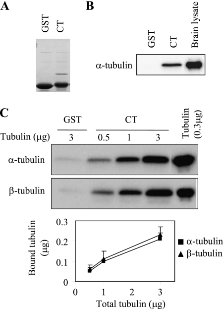
FIGURE 2.
The α2B-AR CT directly interacts with α- and β-tubulin. A, Coomassie Blue staining of GST and GST-α2B-AR CT fusion proteins. B, interaction of the α2B-AR CT with tubulin from rat brain lysates. GST and GST-CT fusion proteins were incubated with rat brain lysates (100 μg) as described under “Experimental Procedures.” Bound α-tubulin was analyzed by Western blotting. C, interaction of the α2B-AR CT with purified tubulin. GST-CT fusion proteins were incubated with increasing amount of purified tubulin (0.5 to 3 μg). Incubation of GST with 3 μg of tubulin was used as a control. Both α- and β-tubulin were analyzed by Western blotting. Bottom panel: quantitative data expressed as the mean ± S.E. of five experiments.

