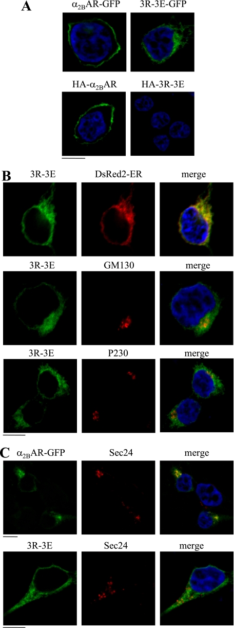
FIGURE 6.
Effect of mutating tubulin binding Arg residues on the subcellular distribution of α2B-AR. A, HEK293 cells cultured on coverslips were transfected with GFP- or HA-tagged α2B-AR or its mutant 3R-3E. The subcellular distribution of the receptors was revealed by detecting GFP fluorescence (upper panel) and detecting HA signal following staining with anti-HA antibodies (lower panel) in non-permeabilized cells as described under “Experimental Procedures.” B, colocalization of the α2B-AR mutant 3R-3E with the ER marker DsRed2-ER, the cis-Golgi marker GM130, and the TGN marker p230. HEK293 cells were transfected with GFP-tagged 3R-3E mutant together with pDsRed2-ER and their co-localization were revealed by fluorescence microscopy, whereas co-localization of the mutant 3R-3E with GM130 and p230 was detected after staining with specific antibody against each marker (1:50 dilution). C, colocalization of α2B-AR and its mutant 3R-3E with the ER exit sites marker Sec24. HEK293 cells were transfected with GFP-tagged α2B-AR plus Sar1H79G (upper panel) or the 3R-3E mutant alone (lower panel). α2B-AR co-localization with Sec24 was revealed after staining with anti-Sec24 antibodies (1:50 dilution). Green, GFP-tagged α2B-AR; red, markers; yellow, co-localization of α2B-AR with the markers; blue, DNA staining by 4,6-diamidino-2-phenylindole (nuclei). The data shown in A, B, and C are representative images of at least three independent experiments. Scale bars, 10 μm.

