FIGURE 5.
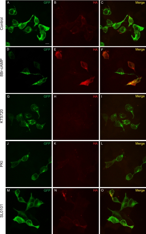
Immunostaining shows PKA activation results in increased surface expression of K2P3.1. Cells transfected with GFP-K2P3.1-HA were grown in the presence of DMSO (control; A–C), 50 μm 8Br-cAMP (D–F), 1 μm KT5720 (G–I), 20 μm PKI (J–L), or 50 μm SL0101 (M–O) and then fixed and stained with anti-HA tag antibody and an Alexa Fluor 647-conjugated secondary antibody. The cells were not permeabilized, so only surface-exposed HA tag in correctly targeted channel was accessible to the antibody. Images are a confocal z-stack. GFP, total expression of the fusion protein; HA, surface-exposed HA tag; Merge, composite image. Scale bar (A), 10 μm.

