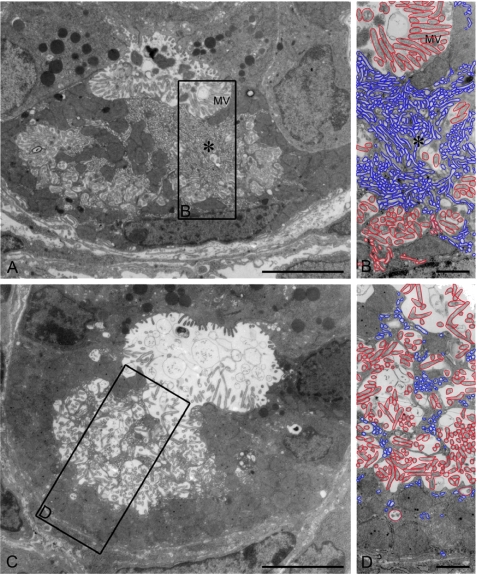
FIGURE 4.
Transmission electron microscopy of stomach mucosa from unstimulated WT oxyntic cells, with some cells more in the resting state (A and B) and some cells in a spontaneously secreting state (C and D). In the resting state, numerous tubulovesicular (tubulocisternal) structures are seen (enlarged in B and D). Scale bars = 5 μm (A and C) and 1 μm (B and D). For better visualization, all tubulocisternal structures are delineated in blue, and all microvillus (MV)-containing secretory membranes are outlined in red.

