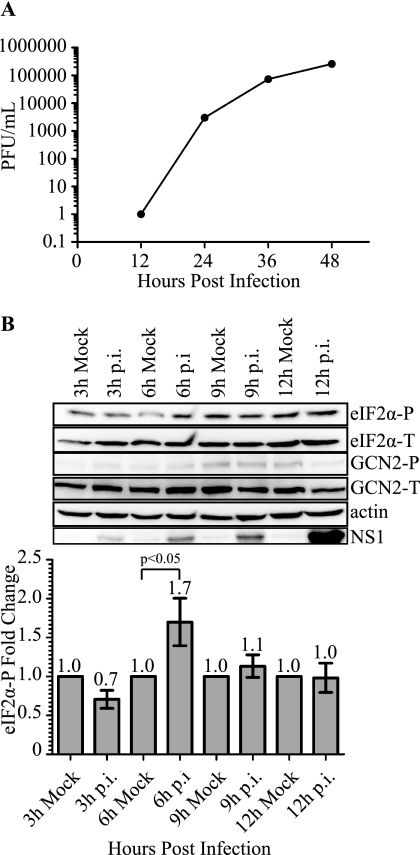
FIGURE 1.
DENV-2 infectivity of 2fTGH cells and induction of eIF2α phosphorylation during infection. A, DENV-2 viral titers as determined by plaque assay. 2fTGH cells were plated overnight and exposed to DENV-2 infection (PL046) at an m.o.i. of 5. Supernatants were collected at the indicated time points post-infection, and infectious virus titer was determined by plaque assay using BHK21 cells. B, phosphorylation state of eIF2α during DENV-2 infection of 2fTGH cells. Mock and DENV-2-infected 2fTGH cell lysates were collected at the indicated time points, and equal amounts of proteins were assayed by Western blot. Phosphorylated eIF2α was visualized using phosphospecific anti-eIF2α antibodies (eIF2α-P) and quantification of eIF2α-P was normalized to total eIF2α (eIF2α-T). Fold-change was measured within each time point as compared with mock infected controls that were assigned a value of 1. Statistical analysis was performed using the Wilcoxon Rank Sum Test (n = 3). Error bars represent the mean ± S.E. GCN2-P was detected using phosphospecific antibodies to GCN2-P, and total GCN2 was visualized with antibodies directed to GCN2. DENV-2 infection was confirmed by immunoblotting for DENV-2 NS1 protein, and actin was used as a loading control. Shown is an immunoblot representative of 3 independent experiments.

