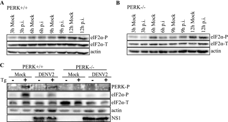
FIGURE 3.
eIF2α phosphorylation during DENV-2 infection in PERK+/+ and PERK−/− cells. A and B, time course of eIF2α phosphorylation in DENV-infected PERK+/+ and PERK−/− MEF cells. Mock and DENV-2-infected (A) PERK+/+ and (B) PERK−/− cell lysates were collected at the indicated time points, and equal amounts of protein were assayed by immunoblot. Levels of eIF2α-P and total eIF2α were visualized as described under “Experimental Procedures.” Actin was included as a loading control. C, DENV-2 inhibits PERK-mediated eIF2α phosphorylation under ER stress. PERK+/+ and PERK−/− cells were plated overnight and infected with DENV-2 (PL046) at an m.o.i. of 5. Three hours p.i. cells were mock-treated or treated with 1 μm Tg for 1 h. Phosphorylated PERK was visualized using phosphospecific PERK antibody, and levels of eIF2α-P and total eIF2α were visualized as described previously. DENV-2 infection was confirmed using a mouse anti-NS1 monoclonal antibody. Actin was used as a loading control.

