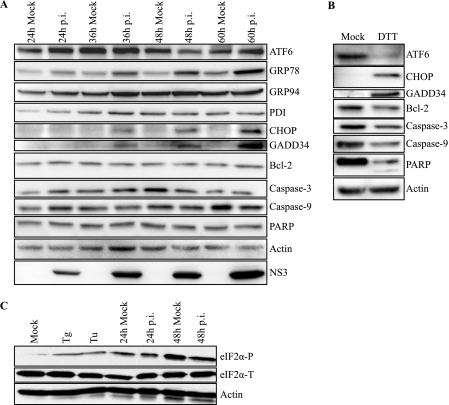
FIGURE 5.
DENV-2 infection induces gene expression of UPR components and activation of the ATF6 pathway. A, immunoblot analysis of components of UPR signaling pathways during a time course of DENV-2 infection. Mock or DENV-2-infected 2fTGH cell lysates were collected at the indicated time points. Lysates were analyzed for the activation and induction of the indicated UPR proteins and markers of apoptosis by immunoblot analysis, as described under “Experimental Procedures.” Actin was used as a loading control, and infection was confirmed by detection of the viral protein NS3. B, positive controls for the induction of UPR proteins and apoptosis markers. Lysates were prepared from 2fTGH cells treated with DTT for 6 h, followed by immunoblot analysis as described under “Experimental Procedures.” C, mock and DENV-2-infected 2fTGH cell lysates were collected at the indicated time points, and equal amounts of proteins were assayed by Western blot. eIF2α-P was visualized and quantified as previously described. Positive controls for eIF2α-P consisted of cells treated with thapsigargin or tunicamycin (Tu) for 30 min. Actin was added as a loading control; the bottom band is the residual HRP activity after probing for eIF2α-P. All data are representative of at least three independent experiments.

