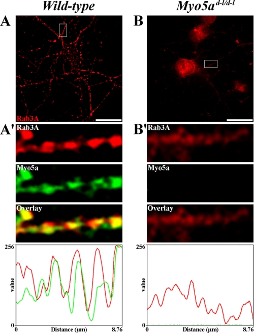
FIGURE 5.
The association of Myo5a with Rab3A is altered in mouse dilute-lethal (Myo5ad-l/d-l) neurons. A–B, indirect immunofluorescent localization of Myo5a (green) and Rab3A (red) in 12 DIV wild-type (A) and 6 DIV dilute-lethal Myo5ad-l/d-l (B) mouse frontal cortex neurons. A′–B′, insets show a high magnification of a dendritic segment where colocalization between Myo5a and Rab3A was detectable in wild-type (A′) but absent in dilute-lethal Myo5ad-l/d-l neurons (B′). Fluorescence intensity was measured for Myo5a (green) and Rab3A (red) along the dendrites shown in the magnified insets (A′–B′). Bars, 20 μm.

