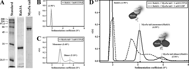
FIGURE 6.
Purification and characterization of the protein-protein interaction between Myo5a tail and Rab3A. A, Myo5a tail and Rab3A were analyzed by SDS-PAGE gel electrophoresis stained with Coomassie after protein purification by affinity and gel filtration chromatography. B–D, examples of diffusion-free sedimentation coefficient distributions (c(s)) derived from sedimentation velocity data of individual Rab3A (2.6 μm, B) and Myo5a tail (0.5 μm, C), and Rab3A plus Myo5a tail together (D). Separate monomer and dimer peaks were observed in the Myo5a tail sample (C). D, GTP-bound Rab3A was found in association with monomeric and dimeric Myo5a tail. The diagrams illustrate possible protein-protein interactions between Myo5a tail regions and GTP-bound Rab3A. All analytical ultracentrifugation experiments were performed in the presence of 1 mm GTPγS or 1 mm GDP.

