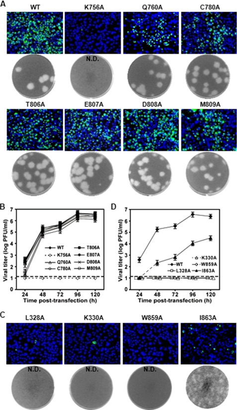
FIGURE 2.
Mutagenesis of cavities A and B using an infectious cDNA clone of DENV-2. A and C, analysis of viral E protein synthesis and plaque morphology for cavity A and B mutant viruses, respectively. BHK-21 cells were transfected with WT and mutant genome-length RNAs (10 μg), and analyzed for viral E protein expression by IFA at 72 h post-transfection (upper panel). Plaque morphologies of WT and mutant viruses were shown (lower panel). N.D., not detectable. B and D, production of cavity A and B mutant viruses after transfection, respectively. Viruses from culture supernatants were collected every 24 h. Viral titers were determined by plaque assay on BHK-21 cells. Error bars indicate the standard deviations from two independent experiments; dashed line, limit of sensitivity of the plaque assay.

